Decoding Tumors: Inside the Spatial Revolution in Cancer Research
Cancer emerges from the very same cellular machinery that keeps us alive. It puzzles both pathologists and the immune system because tumors, unlike infections, share the basic structures and behavioral blueprints of healthy, normal tissue, a resemblance that can let them slip past immune surveillance undetected.
For more than a century, cancer was described by pathologists as a “caricature” of healthy tissue, bizarre distortions of otherwise normal cells.1 But tumors act almost like organisms, evolving within complex, ever-changing ecosystems in which cancerous, immune, and healthy cells all constantly interact and influence one another.2
Until recently, analytic techniques limited scientists’ ability to capture the full complexity of what unfolds within those living systems. Researchers could look at the architecture of a tumor under a microscope or they might break apart lab-grown cancer cells to analyze molecular activity, but they couldn’t do them simultaneously. So those methods were successful, but they ultimately left scientists with fragmented snapshots.
A New Way to See Cancer
Advances in what’s known as spatial transcriptomics have enabled scientists to begin stitching those snapshots together. Now, the landscape of tumors can be mapped in unprecedented detail, revealing how different cells are arranged and interact inside the tissue itself.
This additional level of insight at a cellular level has all sorts of exciting potential applications for patients,” says Dr. Paul Rejto, Head of Discovery Technologies in Pfizer’s Oncology Research Unit. “In terms of what it allows us as researchers to do, the progress is awesome.”
That progress has been made possible by a wave of new technologies that can analyze cancer as it exists in its natural environment, preserving its spatial context.
According to Dr. Rejto, one way of thinking about spatial transcriptomics is as a kind of grid that maps every region of a tumor, all the way down to the single-cell level.
That pinpoint precision matters. Tumors develop when normal cells mutate or defects induce uncontrolled growth, and spatial transcriptomics can be “very, very powerful” for working out where those malfunctions originate, Dr. Rejto says.
“They can start to tell us the exact mechanisms and which specific proteins or genes are defective. That way, we can then learn the causal mechanisms leading to that particular tumor, and how to treat it more effectively.”
A Genomic Foundation
Just a few decades ago, that kind of molecular detail would have been unimaginable.
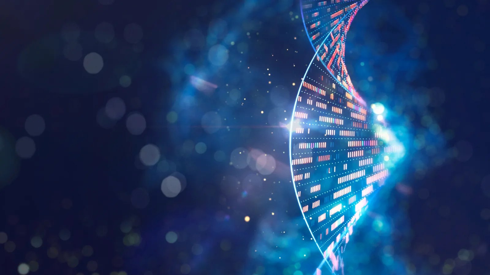
Spatial transcriptomics traces its roots back to the Human Genome Project, the landmark international effort to map the 3 billion pairs of DNA3 that make up the human genome, which completed its mission in 2003.4
It took 13 years and the coordination of 20 different universities and research centers.3 It was an “enormous technological feat,” Dr. Rejto says.
Today, that once-pioneering sequencing is routine. In addition to DNA, spatial transcriptomics can analyze RNA and, more recently, tumor protein profiles. It’s remarkable, Dr. Rejto says, to be able to take the technology that “revolutionized our understanding of human and cancer genetics and now bring that into the next level of spatial understanding.”
Decoding the molecular characteristics of a tumor paves the way for the application of precision medicine that can treat cellular defects with bespoke therapies targeting specific cancer mechanisms.5 And by revealing where immune cells sit relative to a tumor, spatial transcriptomics can also help scientists deduce why certain cancers become resistant to treatment in the first place:
Some tumors are called “cold” because there are very few immune cells nearby. For “immune-excluded” tumors, immune cells are in the vicinity but are held at bay on the tumor’s outskirts, while immune cells can penetrate what are called “infiltrate” tumors.6
Each type poses a unique set of challenges for treatment, making the ability to distinguish between them — something now possible through spatial insight — critical.
The Smoothie Problem
Traditionally, tumors could be studied in two ways. By examining a biopsy under a good old-fashioned microscope, you might spot immune cells, blood vessels, and structural patterns within the tumor. Or, to get deeper genetic information, scientists would take a piece of the tumor, ground it up, and analyze the mixture.
Dr. Rejto likens it to making a smoothie. Examining the blended ingredients can tell you a lot: You might detect bananas, blueberries, and a dash of peanut butter, say. But once everything’s been whizzed up, you lose any sense of how those ingredients were originally arranged.
That situational composition is crucial, though, for treatments such as immunotherapy, which rely on the immune system’s ability to recognize the cancer as foreign. The problem is, “tumors are very creative at masking themselves,” according to Dr. Rejto.
While there are subtle differences, to the immune system, most tumor cells can appear much like normal cells. That’s because they’re largely composed of the same substances. Unlike tumor cells, though, normal cells use the body’s elegant control mechanisms to stop growing when we reach a certain height, say, or know when to regenerate if we cut ourselves. Spatial transcriptomics has been transformative for figuring out when and why the immune system can see beyond a tumor’s disguise.
AI and the Road Ahead
Prior techniques weren’t capable of the same level of molecular insight, but today, spatial transcriptomics takes advantage of similar technology used to make computer chips and “applies it in very clever ways to sequence human DNA,” Dr. Rejto says.
Long stretches of DNA are broken into smaller pieces, analyzed, and assigned a so-called barcode that’s later used to piece back together a high-resolution map of how a tumor is organized.
AI is proving indispensable when deciphering those images, Dr. Rejto says, allowing researchers to identify whether cells are immune or cancerous, gauge their level of aggression, and map how they’re all oriented toward one another.
Combining AI’s formidable aptitude for image analysis, advances in spatial transcriptomics, and the body’s innate ability to regulate cell growth by picking up on the subtleties that differentiate tumors is “miraculous,” Dr. Rejto says. “We can now bring those together to help us take advantage of all the information we're getting.”
Dr. Rejto is particularly excited about how this could all translate into more effective treatments: “The scope of approaches we now have to develop new therapies has exploded.” On the horizon are designer antibodies and more sophisticated early detection methods, “a massively powerful capability” for intervening before cancers evolve, spread, and become more resistant.
“Being able to turn cancer from what it was 25 years ago, a fatal disease, to a chronic disease, to ultimately something where we could actually imagine curing people,” Dr. Rejto says. “That's the dream.”
For the first time, spatial transcriptomics is helping make that dream feel within reach.